We have extensively studied, in the past, effects of mycotoxins on the systemic immune response. We turn now our research to the effects of these toxins on the intestine (first target of toxic food) particularly to the toxicity of mixtures of mycotoxins
3.1. Effects of mycotoxins on systemic immune system
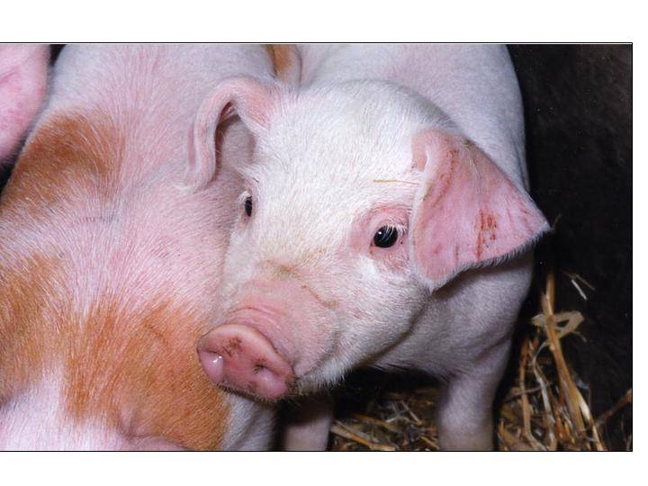
During the last decade, we have investigated the effects of three most problematic mycotoxins (doxynivalenol: DON, Fumonisin B1: FB1 and Aflatoxin B1: AFB1) on the immune response of the piglets. We hypothesized that even when present at low doses, these mycotoxins can alter the immune response, increasing the susceptibility to infectious disease and decreasing the vaccine efficacy.
In piglets, ingestion of FB1-contaminated feed does not alter the total antibody response but significantly decreased the specific antibody response. In vitro, FB1 inhibits, in a dose dependant manner, lymphocyte proliferation (Marin et al., 2007) and modifies cytokine productions (Taranu et al., 2005; Marin et al., 2006). It increases IFN-g synthesis (Th1 cytokine involves in the cellular response) and decreases IL-4 (Th2 cytokine involves in the humoral response). Thus, we proposed that because of its action on lymphocyte proliferation and its ability to drive the cytokine response towards a Th1 response, FB1 decreases the specific antibody response. The ingestion of FB1 was also associated with an increase susceptibility to intestinal and pulmonary infections (Halloy et al., 2005; Devriendt et al., 2010).
A dose dependent experiment indicates that concentration lower than 1mg DON/kg feed had little impact on the immune response of the animals (Accensi et al., 2006). Ingestion of higher doses of DON significantly affects the global and the specific immune response of the pigs. DON does not modulate lymphocytes proliferation after mitogenic stimulation but the toxin had a biphasic effect on lymphocyte proliferation after antigenic stimulation. In the mesenteric lymph node, a significantly lower expression of both TGF-b and IFN-g mRNA expression levels, was observed in animals feed with DON when compared with control piglets (Pinton et al., 2008). At the cellular level, DON was also found to alter the activation of macrophages (Waché et al., 2009) and to impair the functional capacities of neutrophils.
Exposure of piglets to AFB1 delays and decreases the specific lymphocyte proliferation in response to a vaccine antigen, suggesting impaired lymphocyte activation. In these animals, an increased expression of IL-6 in the spleen was observed and in vitro addition of IL-6 decreases the antigenic- but not the mitogenic-induced proliferation of lymphocytes. Because of the effect of IL-6 on the maturation of dendritic cells, we suggest that through its action on IL-6, AFB1 interferes with antigen presentation to lymphocytes and thus reduces their specific proliferative response (Meissonnier et al., 2008).
In conclusion, our studies indicate that mycotoxins, when present at concentration compatible with the one encountered in animal feed, alter the vaccinal immune response of piglets. Depending on the toxin they may act on either the humoral or the cellular immune responses with mechanisms involving their specific action on cytokine synthesis. The effect of mycotoxin on the specific immune response may have implications for humans and animals consuming contaminated food/feed as breakdown in vaccinal immunity may lead to the occurrence of disease even in properly vaccinated populations.
3.2 Effect of mycotoxins on the intestine
The intestine is an immune privileged site where immunoregulatory mechanisms simultaneously defend against pathogens but also preserve tissues homeostasis to avoid immune-mediated pathology in response to environmental challenges. Intestinal epithelium, as the interface between the highly antigenic luminal environment and the mucosal immune system, plays an active role in the immune responsiveness of the intestinal mucosa with the coordinated action of both immune and non-immune cells including dendritic cells, macrophages and epithelial cells. Dendritic cells collaborate with intestinal epithelial cells as sentinels against foreign particulate antigens by building a transepithelial interacting cellular network.
We are interested in the effects of mycotoxins on the intestine in focusing our research on the effects of these toxins on epithelial cells and dendritic cells but also on interactions existing between these two cell types.
§ Effect of mycotoxins on Intestinal Epithelial cells (IECs)
Establishment of the epithelial monolayer by contributing IEC is dependent upon a considerably high degree of intracellular and intercellular organization. Following ingestion of contaminated food or feed, intestinal epithelial cells could be exposed to a high concentration of mycotoxins. The exposure of IEC to these toxins may alter their capacity to proliferate and to insure a proper barrier function. Fusariotoxins are the most detected in temperate north countries. The investigations concerning their effects on the intestinal barrier functions are only beginning. We demonstrated that the fumonisin B1 and the deoxynivalenol both induce a dose-dependent decrease of the Transepithelial electrical resistance (TEER) of a porcine intestinal epithelial cell line. Fumonisin B1 decreases the capacity of epithelial cells to proliferate and to produce IL-8. As this cytokine is involved in neutrophils recruitment, the innate immune response can be affected after fumonisin B1 exposure (Bouhet et al., 2004; Bouhet et al., 2006). For the deoxynivalenol, the observed reduction of TEER was due to an alteration of the tight junction barrier properties as demonstrated by the effect on the permeability to paracellular tracer flux or bacteria (Pinton et al., 2009). The mechanism underlying the trichothecene-induced impairment of intestinal barrier involves the decrease expression of claudins proteins and the MAPK signaling pathway (Pinton et al., 2010).
We now focus on the transcriptomic profile of intestinal tissue in response to DON using intestinal explants treated in vitro with the toxin (Kolf-Clauw et al., 2009) as well in intestinal tissues from DON treated animals (Pinton et al., 2008).
§ Effect of trichothecenes on intestinal dendritic cells
Trichothecene-induced immunosuppression may not be exclusively due to leukocyte apoptosis. Indeed, dendritic cells, that are professional antigen-presenting cells responsible for initiating and suppressing immune responses, are likely mediators for the suppressive effects of trichothecenes on the immune system. DON may also interfere with the maturation and function of intestinal dendritic cells but this aspect of trichothecenes has never been investigated and is the subject of our current research. In these experiments, we are using in monocyte-derived DCs and DCs generated from bone marrow, but the final goal is to repeat these experiments on intestinal DCs. We demonstrated that FLT3-ligand can be use to purified DCs (Guzylack-Piriou et al., 2010).
§ Effect of trichothecenes on the cross talk between intestinal cells
The effects of trichothecenes on the different cell types involved in gut homeostasis can explain various pathologies associated with ingestion of mycotoxins contaminated food. First, DON can potentiate the effect of IL1-b on IL-8 secretion and increase the transepithelial passage of commensal bacteria (Pinton et al., 2009). IL-8 has been implicated in many chronic diseases ranging from inflammatory bowel disease to rheumatoid arthritis. Then, in addition to potentially exacerbate established intestinal inflammation, this mycotoxin may thus participate in induction of sepsis and intestinal inflammation in vivo (Bouhet et Oswald, 2005). Indeed, inflammatory bowel diseases, such as Crohn’s disease, are generally associated with the presence of adherent-invasive bacteria. A hypothesis would be that at least some cases, ingestion of food contaminated with mycotoxins could be involved in inducing inflammatory bowel diseases.
To what extent DON interferes in the cross talk between DC and epithelial cells is the subject of our current research. The ability of intestinal DC to promote gut-tropic lymphocytes is assigned to their unique expression of retinoid hydrogenase enzymes (RALDH) converting dietary vitamin A to retinoic acid (RA), which directly up-regulate domiciliation markers on T cells. Retinoic acid induces a subset of FoxP3+ regulatory T cells, which is important for maintaining immune tolerance in the gut. Therefore, retinoids provide both positive and negative regulatory signals to fine-control the mucosal immune system. We hypothesize that DON may affect the ability of mucosal DCs to interact with intestinal epithelial cells.
3.3 Co-contamination and emerging toxins
Fusarium is the most prevelent fungal species contaminating cereals in temperate regions. Among toxin produces by Fusarium, DON, FB1 and Zearalenone are the most prevalent mycotoxin. However, besides these toxins, Fusarium also produce other toxic secondary metabolites called “emerging” mycotoxins.
With the development of analytical techniques to detect mycotoxins, it has been observed in some raw materials significant amounts of mycotoxins such as fusaproliferin, Beauvericin, moniliformin and enniatins but also the deoxynivalenol-derivates. This raises the interest of better understanding of the potential toxic effects of these “emmergin toxins” and the possible interactions between the different mycotoxins.
The first objective of our study is to characterize the toxic effects of these molecules on gut barrier function using various in vitro models of intestinal epithelial cells and the intestinal explant model. The investigations are focus on the assessment of the cytotoxicity of these toxins, as well as on their effect on the functional properties of the intestinal barrier.
The second objective of our research is to study the effects of co-contamination between major mycotoxins and emerging mycotoxins. It involves determining the type of interaction (additive, synergistic or antagonist). In a first step we have investigated the combined effect of the two major fusariotoxins, DON and FB on pigs. We used The doses used in this trial were very close to the levels naturally found in commodities, and to the limits set by the European Union in feed. The low doses used did not elicit clinical signs but induced microscopic lesions, modulated the immune system and altered the intestine integrity. The effects on the immune responses were only seen when the immune system was activated, following a vaccination protocol in this experiment. Overall, the co-contaminated diet elicited greater effects than the individual diets, especially on liver, on the establishment of the specific immune response, and on proteins involved in the intestinal permeability (Grenier et al., 2010).